References
1. Holly FJ, Lemp MA. Tear physiology and dry eyes. Surv Ophthalmol. 1977;22:69–87.
2. Butovich IA, Millar TJ, Ham BM. Understanding and analyzing meibomian lipids: a review. Curr Eye Res. 2008;33:405–420.
3. Millar TJ, Tragoulias ST , Anderton PJ, et al. The surface activity of purified ocular mucin at the air-liquid interface and interactions with meibomian lipids. Cornea. 2006;25:91–100.
4. King-Smith PE , Hinel EA, Nichols JJ. Application of a novel interferometric method to investigate the relation between lipid layer thickness and tear film thinning. Invest Ophthalmol Vis Sci. 2010;51:2418–2423.
5. Wojtowicz JC, Butovich IA , McCulley JP. Historical brief on composition of human meibum lipids. Ocul Surf. 2009;7:145–153.
6. Butovich IA. The Meibomian puzzle: combining pieces together. Prog Retin Eye Res. 2009;28:483–498.
7. Butovich IA, Uchiyama E, Di Pascuale MA, McCulley JP. Liquid chromatography-mass spectrometric analysis of lipids present in human meibomian gland secretions. Lipids. 2007;42:765–776.
8. Chen J, Green-Church KB, Nichols KK. Shotgun Lipidomic Analysis of Human Meibomian Gland Secretions with Electrospray Ionization Tandem Mass Spectrometry. Invest Ophthalmol Vis Sci. In press.
9. Sullivan BD, Evans JE, Dana MR, Sullivan DA. Influence of aging on the polar and neutral lipid profiles in human meibomian gland secretions. Arch Ophthalmol. 2006;124:1286–1292.
10. Nicolaides N, Kaitaranta JK, Rawdah TN , Macy JI, Boswell FM, 3rd, Smith RE. Meibomian gland studies: comparison of steer and human lipids. Invest Ophthalmol Vis Sci. 1981;20:522–536.
11. Wolff E, ed. The Anatomy of the Eye and Orbit . 4th ed. London : H. K. Lewis and Co; 1954.
12. Holly FJ. Formation and rupture of the tear film. Exp Eye Res. 1973;15:515–525.
13. Shine WE, McCulley JP. Polar lipids in human meibomian gland secretions. Curr Eye Res. 2003;26:89 –94.
14. Butovich IA, Wojtowicz JC, Molai M. Human tear film and meibum: very long chain wax esters and (O-acyl)-omega-hydroxy fatty acids of meibum. J Lipid Res. 2009;50:2471–2485.
15. Butovich IA. Cholesteryl esters as a depot for very long chain fatty acids in human meibum. J Lipid Res. 2009;50:501–513.
16. Saaren-Seppa¨la¨ H, Jauhiainen M, Tervo TM, Redl B, Kinnunen PK, Holopainen JM. Interaction of purified tear lipocalin with lipid membranes. Invest Ophthalmol Vis Sci. 2005;46:3649–3656.
17. Mudgil P, Torres M, Millar TJ. Adsorption of lysozyme to phospholipid and meibomian lipid monolayer films. Colloids Surf B Biointerfaces. 2006;48:128–137.
18. Butovich IA. Lipidomic analysis of human meibum using HPLCMSn. Methods Mol Biol. 2009;579:221–246.
19. Avanti-Polar-Lipids-Inc. Storage and handling of lipids. 2009.
20. Butovich IA, Uchiyama E, McCulley JP. Lipids of human meibum: mass-spectrometric analysis and structural elucidation. J Lipid Res. 2007;48:2220–2235.
21. McCulley JP, Shine WE. Meibomian secretions in chronic blepharitis. Adv Exp Med Biol. 1998;438:319–326.
22. Butovich IA. On the lipid composition of human meibum and tears: comparative analysis of nonpolar lipids. Invest Ophthalmol Vis Sci. 2008;49:3779–3789.
23. Nichols KK, Ham BM, Nichols JJ, Ziegler C, Green-Church KB. Identification of fatty acids and fatty acid amides in human meibomian gland secretions. Invest Ophthalmol Vis Sci. 2007;48:34–39.
24. Borchman D, Foulks GN, Yappert MC, Ho DV. Temperatureinduced conformational changes in human tearlipids hydrocarbon chains. Biopolymers. 2007;87:124–133.
25. Yenice O, Onal S, Midi I, Ozcan E, Temel A, D IG. Visual field analysis in patients with Parkinson's disease. Parkinsonism Relat Disord. 2008;14:193–198.
26. Greiner JV, Glonek T, Korb DR, Booth R, Leahy CD. Phospholipids in meibomian gland secretion. Ophthalmic Res. 1996;28:44–49.
27. Greiner JV, Glonek T, Korb DR, Leahy CD. Meibomian gland phospholipids. Curr Eye Res. 1996;15:371–375.
Kolattukudy PE, Rogers LM, Nicolaides N. Biosynthesis of lipids by bovine meibomian glands. Lipids. 1985;20:468–474.
29. Linton RG, Curnow DH, Riley WJ. The Meibomian glands: an investigation into the secretion and some aspects of the physiology. Br J Ophthalmol. 1961;45:718–723.
30. Jacob JT, Ham B. Compositional profiling and biomarker identification of the tear film. Ocul Surf. 2008;6:175–185.
31. Baron C, Blough HA. Composition of the neutral lipids of bovine meibomian secretions. J Lipid Res. 1976;17:373–376.
32. McCulley JP, Shine W. A compositional based model for the tear film lipid layer. Trans Am Ophthalmol Soc. 1997;95:79–88; discussion 88–93.
33. Shine WE, McCulley JP. The role of cholesterol in chronic blepharitis. Invest Ophthalmol Vis Sci. 1991;32:2272–2280.
34. Sullivan BD, Evans JE, Krenzer KL, Reza Dana M, Sullivan DA. Impact of antiandrogen treatment on the fatty acid profile of neutral lipids in human meibomian gland secretions. J Clin Endocrinol Metab. 2000;85:4866–4873.
35. Ham BM, Jacob JT, Keese MM, Cole RB. Identification, quantification and comparison of major non-polar lipids in normal and dry eye tear lipidomes by electrospray tandem mass spectrometry. J Mass Spectrom. 2004;39:1321–1336.
36. Sullivan BD, Evans JE, Cermak JM, Krenzer KL, Dana MR, Sullivan DA. Complete androgen insensitivity syndrome: effect on human meibomian gland secretions. Arch Ophthalmol. 2002;120:1689–1699.
37. Ham BM, Cole RB, Jacob JT. Identification and comparison of the polar phospholipids in normal and dry eye rabbit tears by MALDITOF mass spectrometry. Invest Ophthalmol Vis Sci. 2006;47: 3330–3338.
38. Ham BM, Jacob JT, Cole RB. MALDI-TOF MS of phosphorylated lipids in biological fluids using immobilized metal affinity chromatography and a solid ionic crystal matrix. Anal Chem. 2005;77: 4439–4447.
39. Silverstein RM, Bassler GC, Morrill TC. Spectrometric Identification of Organic Compounds. 5th ed. New York : John Wiley and Sons; 1991.
40. Skoog DA, Leary JJ. Principles of Instrumental Analysis . 4th ed. New York : Saunders College Publishing; 1992.
41. Borchman D, Foulks GN, Yappert MC, et al. Physical changes in human meibum with age as measured by infrared spectroscopy. Ophthalmic Res. 2010;44:34–42.
42. Borchman D, Foulks GN, Yappert MC, Tang D, Ho DV. Spectroscopic evaluation of human tear lipids. Chem Phys Lipids. 2007; 147:87–102.
43. Oshima Y, Sato H, Zaghloul A, Foulks GN, Yappert MC, Borchman D. Characterization of human meibum lipid using raman spectroscopy. Curr Eye Res. 2009;34:824–835.
44. Foulks GN, Borchman D, Yappert M, Kim SH, McKay JW. Topical azithromycin therapy for meibomian gland dysfunction: clinical response and lipid alterations. Cornea. 2010;29:781–788.
45. Fahy E, Subramaniam S, Brown HA, et al. A comprehensive classification system for lipids. J Lipid Res. 2005;46:839–861.
46. Shevchenko A, Simons K. Lipidomics: Coming to grips with lipid diversity. Nat Rev Mol Cell Biol. 2010;11:593–598.
47. Pes O. Ricerche microchimiche sulla secrezione delle ghiandole sebacee palpebrali. Arch Ottal. 1897;5:82–91.
48. Andrews JS. Human tear film lipids. I. Composition of the principal non-polar component. Exp Eye Res. 1970;10:223–227.
49. Ehlers N. The Precorneal Film. Biomicroscopical, Histological and Chemical Investigations. Acta Ophthalmol Suppl. 1965;(suppl)81: 81–134.
50. Cory CC, Hinks W, Burton JL, Shuster S. Meibomian gland secretion in the red eyes of rosacea. Br J Dermatol. 1973;89:25–27.
51. Nicolaides N. Skin Lipids. Ii. Lipid Class Composition of samples from various species and anatomical sites. J Am Oil Chem Soc. 1965;42:691–702.
52. Tiffany JM. Individual variations in human meibomian lipid composition. Exp Eye Res. 1978;27:289–300.
53. Bron AJ, Tiffany JM. The meibomian glands and tear film lipids; structure, function, and control. Adv Exp Med Biol. 1998;438: 281–295.
54. Bron AJ, Tiffany JM, Gouveia SM, Yokoi N, Voon LW. Functional aspects of the tear film lipid layer. Exp Eye Res. 2004;78:347–360.
55. Ohashi Y, Dogru M, Tsubota K. Laboratory findings in tear fluid analysis. Clin Chim Acta. 2006;369:17–28.
56. Tiffany JM. The lipid secretion of the meibomian glands. Adv Lipid Res. 1987;22:1– 62.
57. Research in dry eye: report of the Research Subcommittee of the International Dry Eye WorkShop. (2007). Ocul Surf. 2007;5:179–193.
58. Keith CG. Seborrhoeic blepharo-kerato-conjunctivitis. Trans Ophthalmol Soc U K. 1967;87:85–103.
59. Harvey DJ, Tiffany JM, Duerden JM, Pandher KS , Mengher LS. Identification by combined gas chromatography-mass spectrometry of constituent long-chain fatty acids and alcohols from the meibomian glands of the rat and a comparison with human meibomian lipids. J Chromatogr. 1987;414:253–263.
60. Nicolaides N, Ruth EC. Unusual fatty acids in the lipids of steer and human meibomian gland excreta. Curr Eye Res. 1982;2:93–98.
61. Nicolaides N, Santos EC. The di- and triesters of the lipids of steer and human meibomian glands. Lipids. 1985;20:454–467.
62. Nicolaides N, Santos EC, Papadakis K. Double-bond patterns of fatty acids and alcohols in steer and human meibomian gland lipids. Lipids. 1984;19:264–277.
63. Nicolaides N, Santos EC, Papadakis K, Ruth EC, Muller L. The occurrence of long chain alpha, omega-diols in the lipids of steer and human meibomian glands. Lipids. 1984;19:990–993.
64. Mathers WD, Lane JA. Meibomian gland lipids, evaporation, and tear film stability. Adv Exp Med Biol. 1998;438:349–360.
65. Dougherty JM, McCulley JP. Analysis of the free fatty acid component of meibomian secretions in chronic blepharitis. Invest Ophthalmol Vis Sci. 1986;27:52–56.
66. Dougherty JM, McCulley JP, Silvany RE, Meyer DR. The role of tetracycline in chronic blepharitis. Inhibition of lipase production in staphylococci. Invest Ophthalmol Vis Sci. 1991;32:2970–2975.
67. Osgood JK, Dougherty JM, McCulley JP. The role of wax and sterol esters of meibomian secretions in chronic blepharitis. Invest Ophthalmol Vis Sci. 1989;30:1958–1961.
68. Shine WE, McCulley JP. Role of wax ester fatty alcohols in chronic blepharitis. Invest Ophthalmol Vis Sci. 1993;34:3515–3521.
69. Shine WE, McCulley JP. Meibomian gland triglyceride fatty acid differences in chronic blepharitis patients. Cornea. 1996;15:340–346.
70. Shine WE, McCulley JP. Meibomianitis: polar lipid abnormalities. Cornea. 2004;23:781–783.
71. Krenzer KL, Dana MR, Ullman MD, et al. Effect of androgen deficiency on the human meibomian gland and ocular surface. J Clin Endocrinol Metab. 2000;85:4874–4882.
72. Butovich IA. Fatty acid composition of cholesteryl esters of human meibomian gland secretions. Steroids. 2010;75:726–733.
73. Joffre C, Souchier M, Gregoire S, et al. Differences in meibomian fatty acid composition in patients with meibomian gland dysfunction and aqueous-deficient dry eye. Br J Ophthalmol. 2008;92: 116–119.
74. Joffre C, Souchier M, Leclere L, et al. Branched-chain fatty acids, increased in tears of blepharitis patients, are not toxic for conjunctival cells. Br J Ophthalmol. 2009;93:1391–1395.
75. McCulley JP, Shine WE. The lipid layer: the outer surface of the ocular surface tear film. Biosci Rep. 2001;21:407–418.
76. McCulley JP, Shine WE. The lipid layer of tears: dependent on meibomian gland function. Exp Eye Res. 2004;78:361–365.
77. Pulfer M, Murphy RC. Electrospray mass spectrometry of phospholipids. Mass Spectrom Rev. 2003;22:332–364.
78. Sommer U, Herscovitz H, Welty FK, Costello CE. LC-MS-based method for the qualitative and quantitative analysis of complex lipid mixtures. J Lipid Res. 2006;47:804–814.
79. Sullivan BD, Evans JE, Dana MR, Sullivan DA. Impact of androgen deficiency on the lipid profiles in human meibomian gland secretions. Adv Exp Med Biol. 2002;506:449–458.
80. Souchier M, Joffre C, Gregoire S, et al. Changes in meibomian fatty acids and clinical signs in patients with meibomian gland dysfunction after minocycline treatment. Br J Ophthalmol. 2008; 92:819–822.
81. Borchman D, Foulks GN, Yappert MC. Human tear lipid compositional, structural and functional relationships using spectroscopy. The Pittsburgh Conference . Chicago , IL ; 2009.
82. Tiffany JM. The meibomian lipids of the rabbit, I: Overall composition. Exp Eye Res. 1979;29:195–202.
83. Tiffany JM, Marsden RG. The meibomian lipids of the rabbit, II; detailed composition of the principal esters. Exp Eye Res. 1982; 34:601–608.
Harvey DJ, Tiffany JM. Identification of meibomian gland lipids by gas chromatography-mass spectrometry: application to the meibomian lipids of the mouse. J Chromatogr. 1984;301:173–187.
85. Harvey DJ. Identification by gas chromatography/mass spectrometry of long-chain fatty acids and alcohols from hamster meibomian glands using picolinyl and nicotinate derivatives. Biomed Chromatogr. 1989;3:251–254.
86. Harvey DJ. Long-chain fatty acids and alcohols from gerbil meibomian lipids. J Chromatogr. 1989;494:23–30.
87. Seyama Y, Otsuka H, Ohashi K, Vivien-Roels B, Pevet P. Sexual dimorphism of lipids in Harderian glands of golden hamsters. J Biochem. 1995;117:661–670.
88. McCulley JP, Dougherty JM, Deneau DG. Classification of chronic blepharitis. Ophthalmology. 1982;89:1173–1180.
89. Shine WE, McCulley JP. Keratoconjunctivitis sicca associated with meibomian secretion polar lipid abnormality. Arch Ophthalmol. 1998;116:849–852.
90. Subbaiah PV, Subramanian VS, Wang K. Novel physiological function of sphingomyelin in plasma: inhibition of lipid peroxidation in low density lipoproteins. J Biol Chem. 1999;274:36409–36414.
91. Augustin AJ, Spitznas M, Kaviani N, et al. Oxidative reactions in the tear fluid of patients suffering from dry eyes. Graefes Arch Clin Exp Ophthalmol. 1995;233:694–698.
92. Shine WE, McCulley JP. Association of meibum oleic acid with meibomian seborrhea. Cornea. 2000;19:72–74.
93. Mathers WD, Stovall D, Lane JA, Zimmerman MB , Johnson S. Menopause and tear function: the influence of prolactin and sex hormones on human tear production. Cornea. 1998;17:353–358.
94. Rolando M, Iester M, Macri A, Calabria G. Low spatial-contrast sensitivity in dry eyes. Cornea. 1998;17:376–379.
95. Gilbard JP, Rossi SR, Heyda KG. Tear film and ocular surface changes after closure of the meibomian gland orifices in the rabbit. Ophthalmology. 1989;96:1180–1186.
96. Goto E, Tseng SC. Differentiation of lipid tear deficiency dry eye by kinetic analysis of tear interference images. Arch Ophthalmol. 2003;121:173–180.
97. Zengin N, Tol H, Gunduz K, Okudan S, Balevi S, Endogru H. Meibomian gland dysfunction and tear film abnormalities in rosacea. Cornea. 1995;14:144–146.
98. Ta CN, Shine WE, McCulley JP, Pandya A, Trattler W, Norbury JW. Effects of minocycline on the ocular flora of patients with acne rosacea or seborrheic blepharitis. Cornea. 2003;22:545–548.
99. US FDA Draft Testing Guidance for Class III Soft Contact Lenses. Washington , DC : US Food and Drug Administration; 1988.
100. US FDA Premarket Notification (510.(k)) for Lens Care Products. Washington , DC : US Food and Drug Administration; 1997.
101. Stone RP. A new perspective for lens care classifying silicone hydrogels (Editorial). June 2007. Available at http://www.siliconehydrogels.org/ editorials/jun_07.asp. Accesssed February 22, 2011.
102. Korb DR , Henriquez AS. Meibomian gland dysfunction and contact lens intolerance. J Am Optom Assoc. 1980;51:243–251.
103. Bontempo AR, Rapp J. Protein-lipid interaction on the surface of a hydrophilic contact lens in vitro. Curr Eye Res. 1997;16:776–781.
104. Prager MD, Quintana RP. Radiochemical studies on contact lens soilation. II. Lens uptake of cholesteryl oleate and dioleoyl phosphatidylcholine. J Biomed Mater Res. 1997;37:207–211.
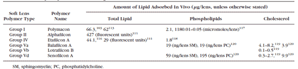
105. Ho CH, Hlady V. Fluorescence assay for measuring lipid deposits on contact lens surfaces. Biomaterials. 1995;16:479–482.
106. Lord MS, Stenzel MH, Simmons A, Milthorpe BK. The effect of charged groups on protein interactions with poly(HEMA) hydrogels. Biomaterials. 2006;27:567–575.
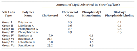
107. Carney FP, Nash WL, Sentell KB. The adsorption of major tear film lipids in vitro to various silicone hydrogels over time. Invest Ophthalmol Vis Sci. 2008;49:120–124.
108. Iwata M, Ohno S, Kawai T, Ichijima H, Cavanagh HD. In vitro evaluation of lipids adsorbed on silicone hydrogel contact lenses using a new gas chromatography/mass spectrometry analytical method. Eye Contact Lens. 2008;34:272–280.
109. Hart DE. Lipid deposits which form on extended wear contact lenses. International Contact Lens Clinics. 1984;11:348–360.
110. Hart DE, Tidsale RR, Sack RA. Origin and composition of lipid deposits on soft contact lenses. Ophthalmology. 1986;93:495–503.
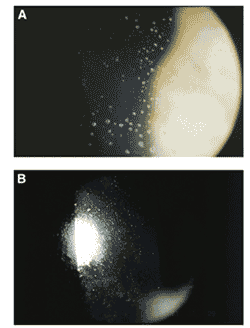
111. Bowers RW, Tighe BJ. Studies of the ocular compatibility of hydrogels: white spot deposits—chemical composition and geological arrangement of components. Biomaterials. 1987;8:172–176.
112. Bowers RW, Tighe BJ. Studies of the ocular compatibility of hydrogels; white spot deposits—incidence of occurrence, location and gross morphology. Biomaterials. 1987;8:89 –93.
113. Hart DE, Lane BC, Josephson JE, et al. Spoilage of hydrogel contact lenses by lipid deposits: tear-film potassium depression, fat, protein, and alcohol consumption. Ophthalmology. 1987;94: 1315–1321.
114. Bontempo AR, Rapp J. Lipid deposits on hydrophilic and rigid gas permeable contact lenses. CLAO J. 1994;20:242–245.
115. Franklin V, Horne A, Jones L, Tighe B. Early deposition trends on group I (Polymacon and Tetrafilcon A) and group III (Bufilcon A) materials. CLAO J. 1991;17:244–248.
116. Bontempo AR, Rapp J. Protein and lipid deposition onto hydrophilic contact lenses in vivo. CLAO J. 2001;27:75–80.
117. Jones L, Fcoptom, Mann A, Evans K, Franklin V, Tighe B. An in vivo comparison of the kinetics of protein and lipid deposition on group II and group IV frequent-replacement contact lenses. Optom Vis Sci. 2000;77:503–510.
118. Jones L, Evans K, Sariri R, Franklin V, Tighe B. Lipid and protein deposition of N-vinyl pyrrolidone-containing group II and group IV frequent replacement contact lenses. CLAO J. 1997;23:122–126.
119. Zhao Z, Carnt NA, Aliwarga Y, et al. Care regimen and lens material influence on silicone hydrogel contact lens deposition. Optom Vis Sci. 2009;86:251–259.
120. Cheung SW, Cho P, Chan B, Choy C, Ng V. A comparative study of biweekly disposable contact lenses: silicone hydrogel versus hydrogel. Clin Exp Optom. 2007;90:124–131.
121. Jones L, Franklin V, Evans K, Sariri R, Tighe B. Spoilation and clinical performance of monthly vs. three monthly group II disposable contact lenses. Optom Vis Sci. 1996;73:16 –21.
122. Rapp J, Broich JR. Lipid deposits on worn soft contact lenses. CLAO J. 1984;10:235–239.
123. Jones L, Senchyna M, Glasier MA, et al. Lysozyme and lipid deposition on silicone hydrogel contact lens materials. Eye Contact Lens. 2003;29:S75–79; discussion S83–S74, S192–S194.
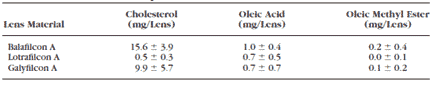
124. Maziarz EP, Stachowski MJ, Liu XM, et al. Lipid deposition on silicone hydrogel lenses, Part I: quantification of oleic acid, oleic acid methyl ester, and cholesterol. Eye Contact Lens. 2006;32: 300–307.
125. Morris CA, Holden BA, Papas E, et al. The ocular surface, the tear film, and the wettability of contact lenses. Adv Exp Med Biol. 1998;438:717–722.
126. Young WH, Hill RM. Tear cholesterol levels and contact lens adaptation. Am J Optom Arch Am Acad Optom. 1973;50:12–16.
127. Yamada M, Mochizuki H, Kawashima M, Hata S. Phospholipids and their degrading enzyme in the tears of soft contact lens wearers. Cornea. 2006;25:S68 –72.
128. Kawashima M, Yamada M, Arita R, Kawai M, Mashima Y. Measurement of phospholipids in human tears. Folia Ophthalmol Jpn. 2003;54:870–873.
129. Dumbleton KA, Woods CA, Jones LW, Fonn D. Comfort and adaptation to silicone hydrogel lenses for daily wear. Eye Contact Lens. 2008;34:215–223.
130. Fonn D. Targeting contact lens induced dryness and discomfort: what properties will make lenses more comfortable. Optom Vis Sci. 2007;84:279–285.
131. Guillon JP, Morris J, Hall B. Evaluation of the pre-lens tear film forming on three disposable contact lenses. Adv Exp Med Biol. 2002;506:901–915.
132. Maissa C, Guillon M, Girard-Claudon K, Cooper P. Tear lipid composition of hydrogel contact lens wearers. Adv Exp Med Biol. 2002;506:935–938.
133. Hume EB, Cole N, Parmar A, et al. Secretory phospholipase A2 deposition on contact lenses and its effect on bacterial adhesion. Invest Ophthalmol Vis Sci. 2004;45:3161–3164.
134. Mochizuki H, Yamada M, Hatou S, Kawashima M, Hata S. Deposition of lipid, protein, and secretory phospholipase A2 on hydrophilic contact lenses. Eye Contact Lens. 2008;34:46–49.
135. Guillon JP. Tear Film Structure and Contact Lenses . Lubbock , TX : Dry Eye Institute; 1986.
Korb DR. Tear film-contact lens interactions. Adv Exp Med Biol. 1994;350:403–410.
137. Korb DR, Greiner JV, Glonek T. Tear film lipid layer formation: implications for contact lens wear. Optom Vis Sci. 1996;73:189–192.
138. Young G, Efron N. Characteristics of the pre-lens tear film during hydrogel contact lens wear. Ophthalmic Physiol Opt. 1991;11:53–58.
139. Guillon M, Guillon JP. Hydrogel lens wettability during overnight wear. Ophthalmic Physiol Opt. 1989;9:355–359.
140. Thai LC, Tomlinson A, Doane MG. Effect of contact lens materials on tear physiology. Optom Vis Sci. 2004;81:194–204.
141. Guillon M, Maissa C. Use of silicone hydrogel material for daily wear. Cont Lens Anterior Eye. 2007;30:5–10; quiz 71.
142. Guillon M, Styles E, Guillon JP, Maissa C. Preocular tear film characteristics of nonwearers and soft contact lens wearers. Optom Vis Sci. 1997;74:273–279.
143. Glasson MJ, Stapleton F, Keay L, Sweeney D, Willcox MD. Differences in clinical parameters and tear film of tolerant and intolerant contact lens wearers. Invest Ophthalmol Vis Sci. 2003;44: 5116–5124.
144. Glasson M, Stapleton F, Willcox M. Lipid, lipase and lipocalin differences between tolerant and intolerant contact lens wearers. Curr Eye Res. 2002;25:227–235.
145. Gasymov OK, Abduragimov AR , Yusifov TN , Glasgow BJ. Binding studies of tear lipocalin: the role of the conserved tryptophan in maintaining structure, stability and ligand affinity. Biochim Biophys Acta. 1999;1433:307–320.
146. Glasgow BJ, Abduragimov AR , Farahbakhsh ZT, Faull KF, Hubbell WL. Tear lipocalins bind a broad array of lipid ligands. Curr Eye Res. 1995;14:363–372.
147. Fullard RJ. Identification of proteins in small tear volumes with and without size exclusion HPLC fractionation. Curr Eye Res. 1988;7:163–179.
148. Tsai PS, Evans JE, Green KM, et al. Proteomic analysis of human meibomian gland secretions. Br J Ophthalmol. 2006;90:372–377.
149. Glasgow BJ, Abduragimov AR , Gasymov OK, Yusifov TN. Tear lipocalin: structure, function and molecular mechanisms of action. Adv Exp Med Biol. 2002;506:555–565.
150. Redi B. Human tear lipocalin. Biochim Biophys Acta. 2000; 1482:241–248.
151. Glasgow BJ, Gasymov OK. Focus on molecules: tear lipocalin. Exp Eye Res. 2010;Aug 21 [ePub ahead of print].
152. Gasymov OK, Abduragimov AR , Yusifov TN , Glasgow BJ. Structural changes in human tear lipocalins associated with lipid binding. Biochim Biophys Acta. 1998;1386:145–156.
153. Glasgow BJ, Marshall G, Gasymov OK, Abduragimov AR , Yusifov TN, Knobler CM. Tear lipocalins: potential lipid scavengers for the corneal surface. Invest Ophthalmol Vis Sci. 1999;40:3100–3107.
154. Gasymov OK, Abduragimov AR , Glasgow BJ. Intracavitary ligand distribution in tear lipocalin by site-directed tryptophan fluorescence. Biochemistry. 2009;48:7219–7228.
155. Breustedt DA, Chatwell L, Skerra A. A new crystal form of human tear lipocalin reveals high flexibility in the loop region and induced fit in the ligand cavity. Acta Crystallogr D Biol Crystallogr. 2009;65:1118–1125.
156. Gouveia SM, Tiffany JM. Human tear viscosity: an interactive role for proteins and lipids. Biochim Biophys Acta. 2005;1753:155–163.
157. Gasymov OK, Abduragimov AR , Glasgow BJ. Evidence for internal and external binding sites on human tear lipocalin. Arch Biochem Biophys. 2007;468:15–21.
158. Yamada M, Mochizuki H, Kawai M, Tsubota K, Bryce TJ. Decreased tear lipocalin concentration in patients with meibomian gland dysfunction. Br J Ophthalmol. 2005;89:803–805.
159. Millar TJ, Mudgil P, Butovich IA , Palaniappan CK. Adsorption of human tear lipocalin to human meibomian lipid films. Invest Ophthalmol Vis Sci. 2009;50:140–151.
160. Mateu L, Caron F, Luzzati V, Billecocq A. The influence of proteinlipid interactions on the order-disorder conformational transitions of the hydrocarbon chain. Biochim Biophys Acta. 1978; 508:109–121.
161. Miano F, Calcara M, Millar TJ, Enea V. Insertion of tear proteins into a meibomian lipids film. Colloids Surf B Biointerfaces. 2005; 44:49 –55.
162. Tragoulias ST , Anderton PJ, Dennis GR, Miano F, Millar TJ. Surface pressure measurements of human tears and individual tear film components indicate that proteins are major contributors to the surface pressure. Cornea. 2005;24:189–200.
163. Holzfeind P, Merschak P, Dieplinger H, Redl B. The human lacrimal gland synthesizes apolipoprotein D mRNA in addition to tear prealbumin mRNA, both species encoding members of the lipocalin superfamily. Exp Eye Res. 1995;61:495–500.
164. Jauhiainen M, Setala NL, Ehnholm C, et al. Phospholipid transfer protein is present in human tear fluid. Biochemistry. 2005;44: 8111–8116.
165. Tiffany JM. Tears in health and disease. Eye ( Lond ) . 2003;17:923–926.
166. Perkins RE, Kundsin RB, Pratt MV, Abrahamsen I, Leibowitz HM. Bacteriology of normal and infected conjunctiva. J Clin Microbiol. 1975;1:147–149.
167. Stapleton F, Willcox MD, Fleming CM, Hickson S, Sweeney DF, Holden BA. Changes to the ocular biota with time in extendedand daily-wear disposable contact lens use. Infect Immun. 1995; 63:4501–4505.
168. Dougherty JM, McCulley JP. Bacterial lipases and chronic blepharitis. Invest Ophthalmol Vis Sci. 1986;27:486–491.
169. McCulley JP, Dougherty JM. Bacterial aspects of chronic blepharitis. Trans Ophthalmol Soc U K. 1986;105(Pt 3):314 –318.
170. Ridsdale RA, Palaniyar N, Possmayer F, Harauz G. Formation of folds and vesicles by dipalmitoylphosphatidylcholine monolayers spread in excess. J Membr Biol. 2001;180:21–32.
171. Akiyama J, Hoffman A, Brown C, et al. Tissue distribution of surfactant proteins A and D in the mouse. J Histochem Cytochem. 2002;50:993–996.
172. Madsen J, Kliem A, Tornoe I, Skjodt K, Koch C, Holmskov U. Localization of lung surfactant protein D on mucosal surfaces in human tissues. J Immunol. 2000;164:5866–5870.
173. Stahlman MT, Gray ME, Hull WM, Whitsett JA. Immunolocalization of surfactant protein-D (SP-D) in human fetal, newborn, and adult tissues. J Histochem Cytochem. 2002;50:651–660.
174. Ni M, Evans DJ, Hawgood S, Anders EM, Sack RA, Fleiszig SM. Surfactant protein D is present in human tear fluid and the cornea and inhibits epithelial cell invasion by Pseudomonas aeruginosa. Infect Immun. 2005;73:2147–2156.
175. Brauer L, Johl M, Borgermann J, Pleyer U, Tsokos M, Paulsen FP. Detection and localization of the hydrophobic surfactant proteins B and C in human tear fluid and the human lacrimal system. Curr Eye Res. 2007;32:931–938.
176. Brauer L, Kindler C, Jager K, et al. Detection of surfactant proteins A and D in human tear fluid and the human lacrimal system. Invest Ophthalmol Vis Sci. 2007;48:3945–3953.
177. Pfister RH, Soll RF. New synthetic surfactants: the next generation? Biol Neonate. 2005;87:338–344.
178. Sorensen GL, Husby S, Holmskov U. Surfactant protein A and surfactant protein D variation in pulmonary disease. Immunobiology. 2007;212:381–416. |